


Hospitals
Specialties & Packages
Our Services
Health Hub
Patients & Visitors
Partner
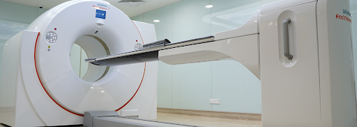
PET is an advanced nuclear medical imaging test that can give a thorough insight into the functions of an organ or system in your body. PET scans are more often carried out to give diagnosis of cancers, neurological (brain) disorders, and cardiovascular (heart) diseases.
During the Positron Emission Tomography scan procedure, a radioactive tracer is injected, and images of your body are being recorded. A camera detects the emissions from the injected radioactive tracer and forms multi-dimensional images that are computer-generated. Generally, the injected radiotracers accumulate in diseased tissues more than in healthier tissues.
There are more PET scanners being put together with Computed Tomography (CT) scans. This enables the information gathered by CT images to be put along with the PET's functional data.
Essentially, PET scans are for providing accurate diagnosis and to monitor conditions that include cancers, neurological (brain) disorders, and cardiovascular (heart-related) diseases. In the early stages, these diseases can be detected by using other imaging tests (such as CT and MRI scans). Mainly, it helps your doctor to determine which stage cancer cells are in, where and how much they have spread to various parts of the body and what the cancer treatment will be.
A PET scan will be able to contribute to determining whether surgery will be necessary for epileptic seizures by providing information on which part of the brain is responsible for your epilepsy. PET scan images are also able to help determine the areas where the brain does not function normally, thus it is beneficial for monitoring Alzheimer's Disease and Parkinson's. Detecting neurological disorders in their early stages can lead to more effective treatment.
After undergoing a PET scan, most people do not experience any significant side effects. The radioactive material used in the PET scanner quickly loses its activity, and it is eliminated from the body through urine or faeces. In addition, the dose of radiation is very small — similar to several years' worth of natural radiation from the environment. Allergic reactions are very rare and usually minor. As a result, there is typically no need for any special precautions or restrictions after the procedure, and you can usually go home the same day after taking off your hospital gown.

Wait a minute