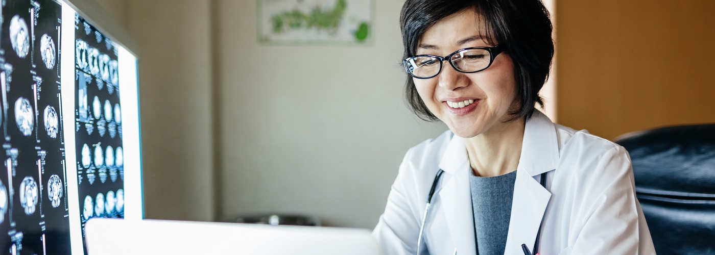
Radiology is a specialised branch of medicine that focuses on the use of imaging technology to view the inside of the body for diagnostic purposes. Radiologists are medical doctors specialising in the use of advanced equipment and techniques to diagnose, treat, and manage various medical conditions, ensuring that patients receive accurate and timely care.
Gleneagles Hospital Kuala Lumpur boasts ultramodern radiological and imaging equipment with state-of-the-art technology and is managed by a dedicated team of specialist radiologists and technologists led by a highly qualified and experienced senior radiological consultant.
For concerned mothers, we also offer non-invasive tests that detect major chromosomal issues through maternal blood evaluation, including prenatal screening with biochemical markers and risk evaluation.
Gleneagles Hospital Kuala Lumpur offers comprehensive perinatal radiological screening services for expectant mothers, ensuring the health of both mother and child. Our services include:
Our radiology department operates with advanced RIS (Radiology Information System) and PACS (Picture Archiving Communication System). Once the investigation has concluded, images are immediately available to both radiologists and treating physicians via computer system. This digital process enables quicker interpretation, allowing radiological specialists to assess the results without delay. By utilising RIS and PACS, we minimise the risk of losing important medical images and reports, thereby providing a more secure and efficient workflow for patient care.
With PACS, all imaging tests and radiology reports are digitally stored and accessible at any time, allowing for easy comparison with previous results. This capability helps our radiology specialists provide consistent and high-quality care across all your imaging services.
Gleneagles Hospital Kuala Lumpur has recently upgraded its CT Scan diagnostic technology to offer multi-slice and multi-formatted 3D imaging, enhancing our diagnostic capabilities. This advanced technology allows us to conduct scans in seconds, providing highly detailed images of the brain, spinal cord, and coronary arteries, among other critical areas. It is particularly useful for non-invasive procedures, such as calcium scoring estimation for coronary arteries, which is becoming increasingly popular with both patients and radiology specialists.
Our CT-guided biopsies and routine drainage procedures benefit from the latest pressure injector technology, ensuring precise injections of contrast medium for clear imaging results. Each 3D image created provides excellent help with the evaluation of issues such as fractures, tumors, and aneurysms with great accuracy.
For complex conditions like pulmonary embolisms, issues with abdominal arteries, and extremities, including neck and brain vessels, our CT angiogram provides a minimally invasive solution, offering guided drainage procedures and significantly reducing the need for more invasive surgeries.
Cardiac MRI is a non-invasive diagnostic imaging technique that provides detailed insights into various cardiovascular conditions. This advanced imaging tool is crucial for diagnosing and evaluating:
Unlike traditional MRI, Cardiac MRI is specifically optimised for the cardiovascular system, allowing for more accurate assessment of key functions and morphological features. This optimization utilizes ECG gating and rapid imaging techniques, enabling faster and more precise results.
Some of the benefits of Cardiac MRI include:
MRI (3T) represents the latest advancement in Magnetic Resonance Imaging technology, providing exceptional accuracy in diagnosing conditions affecting the brain, spine, joints, and pelvic organs. The 3T MRI is also capable of performing detailed imaging of the liver, biliary tree (MRCPs) as well as pelvic organs, ensuring comprehensive diagnostic coverage.
Recent developments and progress in MRI technology have enhanced its capabilities to conduct breast imaging via a special breast coil specially created for cases that pose diagnostic difficulty. Additionally, the 3T MRI now allows for high-resolution scans of the arteries and veins of the brain, body, abdomen, and other extremities, offering detailed insights into vascular health.
With its superior imaging capabilities, 3T MRI allows our specialists to deliver the most accurate diagnoses for a wide range of neurological, musculoskeletal, and vascular conditions.
At Gleneagles Hospital Kuala Lumpur, we offer advanced radiology services to help diagnose and treat a wide range of conditions. From CT scans and MRI (3T) to cardiac MRI diagnostics and perinatal screenings, our state-of-the-art imaging technologies provide accurate and timely results. Whether you're experiencing symptoms or undergoing routine health checks, the team of expert radiologists and specialists is here to provide the best care tailored to your needs.
Book your consultation today at Gleneagles Hospital Kuala Lumpur and benefit from the most advanced medical imaging techniques available. We are committed to ensuring your health with the highest quality diagnostic services, all in a safe and caring environment.
With expertise in CT scans, MRI (3T), cardiac MRI diagnostics, and perinatal screenings, the top radiologists at Gleneagles Hospital Kuala Lumpur are here to provide exceptional care for your diagnostic needs. Choose from the following experienced specialists, each offering personalised care and advanced imaging technology tailored to your unique health requirements. Book a consultation today and meet the best radiologists near you for expert diagnostic imaging services.


Wait a minute

Wait a minute