The heart is a complex organ made up of chambers, valves and blood vessels that work together to pump blood throughout the body. While standard heart imaging techniques provide valuable information, some conditions require more detailed visualisation to accurately diagnose and plan treatment.
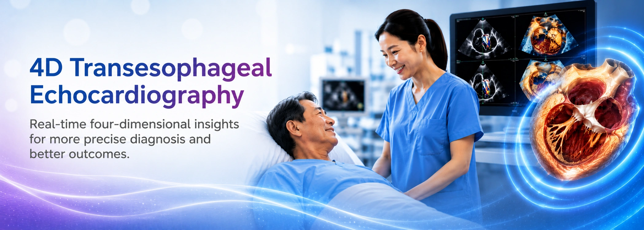
Advances in cardiac imaging have made it possible for doctors to assess heart structures with greater precision. One such technology is 4D Transoesophageal Echocardiography (4D-TEE), an advanced imaging technique that provides highly detailed, real-time, three-dimensional views of the heart.
By delivering clearer visualisation of the heart's anatomy and function, 4D-TEE plays an important role in diagnosing complex heart conditions and guiding both surgical and minimally invasive cardiac procedures.
4D Transoesophageal Echocardiography (4D-TEE) is an advanced form of cardiac ultrasound that uses a specialised ultrasound probe placed gently into the oesophagus to obtain detailed images of the heart.
Because the oesophagus sits directly behind the heart, the probe is able to capture high-resolution images without interference from the chest wall or lungs.
Unlike conventional echocardiography, 4D-TEE produces real-time, three-dimensional images that allow doctors to assess the heart from multiple angles while it is beating. This provides a more comprehensive understanding of the heart's structure, valve function and blood flow.
Your cardiologist may recommend a 4D-TEE when more detailed heart imaging is required to diagnose or evaluate certain cardiac conditions.
4D-TEE is commonly used for:
Not every patient requires a 4D-TEE examination. Your cardiologist will recommend the most appropriate imaging test based on your symptoms, medical history and overall clinical assessment.
Before the procedure begins, patients are usually asked to fast for several hours. A local anaesthetic spray is applied to numb the throat, and sedation may be given to help ensure comfort throughout the examination.
A thin, flexible ultrasound probe is then gently passed through the mouth into the oesophagus. From this position, detailed ultrasound images of the heart are captured in real time while the heart continues to beat naturally.
The examination typically takes between 20 and 60 minutes, depending on the complexity of the assessment. Once completed, patients are monitored until the effects of the sedation have worn off before returning home.
4D Transoesophageal Echocardiography offers several important advantages compared to conventional heart imaging techniques, including:
Many heart conditions may not cause obvious symptoms during their early stages. Accurate cardiac imaging allows doctors to detect abnormalities sooner, evaluate their severity and recommend the most appropriate treatment.
Advanced imaging technologies such as 4D-TEE help cardiologists make well-informed clinical decisions while improving procedural planning for patients undergoing structural heart interventions or heart valve treatment.
Early and accurate diagnosis also provides greater reassurance, allowing patients to better understand their condition and receive timely care when needed.
Modern cardiac care combines specialist expertise with advanced imaging technology to improve diagnosis, treatment planning and patient outcomes.
At Gleneagles Hospital Kuala Lumpur, advanced cardiac imaging, including 4D Transoesophageal Echocardiography (4D-TEE), supports comprehensive evaluation for patients with complex heart conditions and those requiring structural heart interventions.
https://gleneagles.com.my/kuala-lumpur/medical-specialties/cardiology

Wait a minute
Wait a minute
 CHI
CHI
 e-Shop
 找医生
找医生
 请求预约
请求预约
 国际患者
国际患者
 +603 4141 3000
 +603 4141 3131