What Is Spinal Stenosis?
Spinal stenosis is the narrowing of the spinal canal (spaces within the spine) that may exert pressure on the nerves along the spine. Spinal stenosis occurs most often in the lower back and the neck.
What Causes Spinal Stenosis?
The main cause of spinal stenosis is bone degeneration due to ageing. As we grow older, the vertebrae and intervertebral discs are prone to collapse due to degeneration. The collapsed vertebrae may compress and cause the rupture and inflammation of the intervertebral disc. Chronic inflammation of the small joints also causes the bones to grow spurs that oppress the spinal cord and spinal nerve roots, which leads to spinal stenosis.
Other causes of spinal stenosis include:
- Tumour
- Bone fragments due to fractures
- Abscess
- Scoliosis due to old-age degeneration
- Thickening of the ligaments in of the spine
Symptoms
Some patients with spinal stenosis may not have any symptoms, while other patients may have multiple symptoms. In the case of spinal stenosis, symptoms may worsen over time. Symptoms may vary depending on the location of the stenosis and which nerves are affected.
- Leg pain
- Leg paralysis
- Numbness in the leg
- Weakness in the leg
- Inability to walk
- Immobility
Diagnosis
To diagnose spinal stenosis, your doctor may ask you about signs and symptoms, discuss your medical history, and conduct a physical examination.
Your doctor may order several imaging tests to help pinpoint the cause of your signs and symptoms. Some of the tests include:
Who Are At Risk?
There are certain risk factors that may lead to spinal stenosis. Some of the risk factors include:
- Elderly people
- Workers who have long been burdened with heavy work due to work needs
- Those who have long-term bad sitting postures
- Those with family members suffering from spinal stenosis
Treatment
The treatment of spinal stenosis consists of conservative treatment and surgical treatment. Conservative treatment is more suitable for patients with mild symptoms. Patients with severe symptoms affecting quality of life should consider surgery to solve the root cause of their condition.
- Conservative treatment:
- Medication and physical therapy – taking analgesic, anti-inflammatory or swell-reducing medication combined with physical therapy can reduce the swelling of the spinal nerve roots, thus reducing pain.
- Steroid injection / infiltration – Steroids can reduce inflammation and swelling that cause the spinal nerve roots to be oppressed. However, the steroid injection only reduces pain temporarily and does not solve the root cause of spinal stenosis.
- Radiofrequency modulation – A needle-sized medical device is inserted into the lumbar spine to eliminate the pain at the nerve. This method is more effective than steroid injection / infiltration and provides longer relief.
- Surgical treatment:
- Decompression – This surgery aims to remove the roots of the spinal nerve roots. If the spinal stenosis is caused by bone spurs, spur resection will be done; If it is caused by thickening of the ligamentum flavum, the ligamentum flavum will be removed. The benefit of decompression surgery is that this is a low-risk procedure and the flexibility of the lumbar spine is maintained after surgery. Decompression surgery can be performed via mini-open technique, video-assisted tubular system, or endoscopic technique.
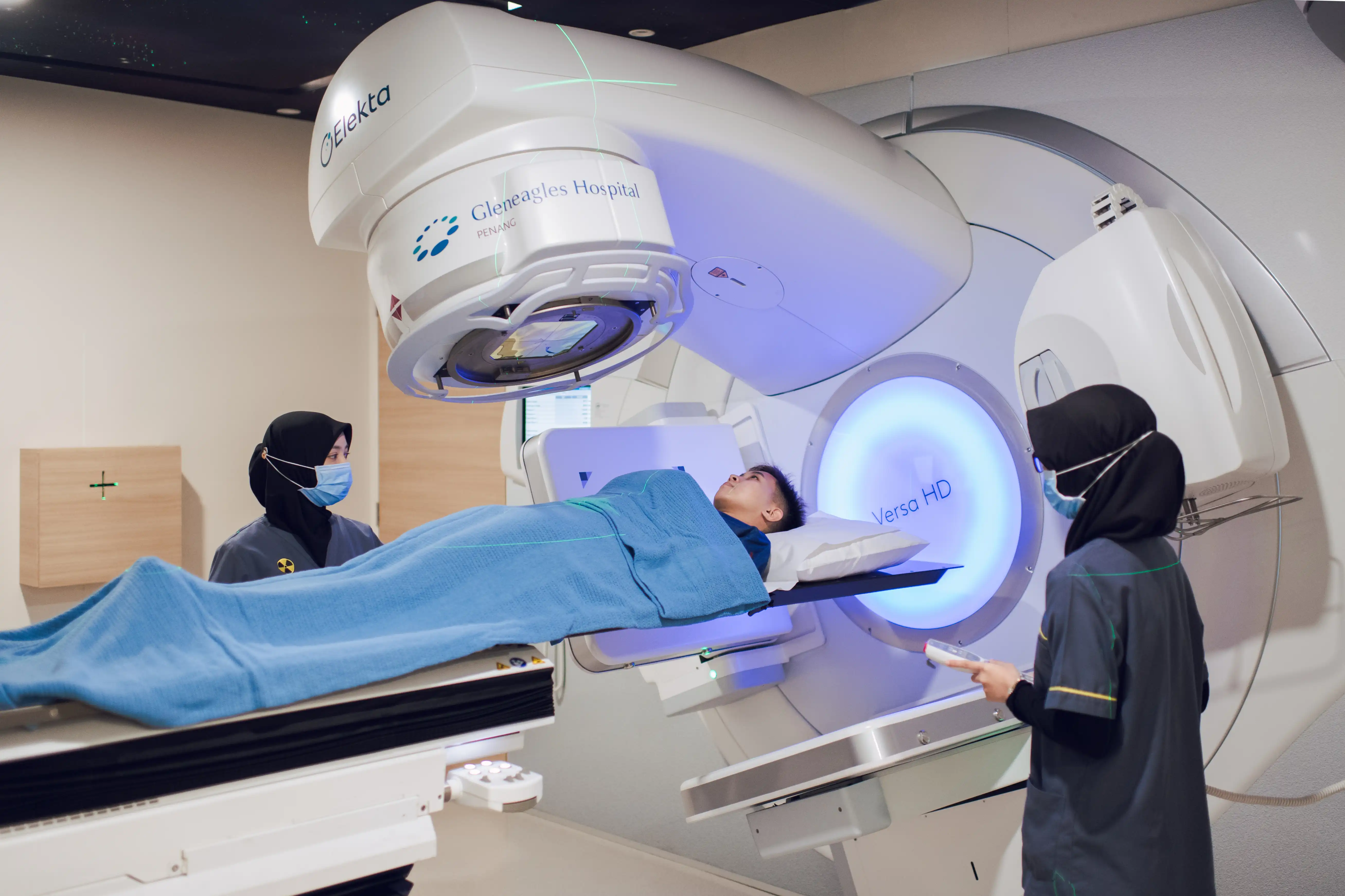
Endoscopic spine surgery (ESS) is an advanced form of minimally invasive spine surgery designed to provide the patient a quicker recovery time and less recurring pain than traditional spine surgery methods. ESS also can help preserve normal range of spine mobility post-operatively.
In the experienced hands of a spine surgeon who regularly performs endoscopic spine surgery using the endoscope—the surgery is performed in a different way offering patients many potential benefits, including:
i. Endoscopes reduce the need to cut through soft tissues (eg, skin tissue-to-muscle injury or damage)
ii. Less blood loss
iii. Less post-operative discomfort or pain
iv. Fast recovery and healing
• Spinal fusion – This surgery aims to fixate the two vertebral bodies by implanting screws or cages across the spinal joints, removing the original intervertebral discs and fusing the joints. Recent advances have made percutaneous screw fixation possible as part of the minimally invasive surgery (MIS).

i. TLIF - Transforaminal Lumbar Interbody Fusion
TLIF is a form of spine surgery in which the lumbar spine is approached through an incision in the back Fusion is a means of stabilizing the spine by fusing two vertebrae together. The transforaminal approach accesses the disc through the space between the vertebrae. This technique allows the surgeon to gently separate the muscles surrounding the spine instead of cutting through them.
ii. OLIF - Oblique lumbar interbody fusion
Accesses and repairs the lower (lumbar) spine from the front and side of the body (passing in a trajectory about halfway between the middle of the stomach and the side of the body). By applying an oblique lateral approach to the spine, the procedure enable placement of a large interbody graft on to the apophysis, the strongest part of a vertebral body. The graft provides anterior column support while avoiding risks and obstacles associated with traditional anterior, posterior, and direct lateral approaches.
iii. ALIF - Anterior Lumbar Interbody Fusion
This is a spinal operation where the spine is approached from the front rather than from the back. It is performed through the abdomen. The disc is removed and pressure on the nerves can be relieved. A large cage is then placed in the space where the disc was located, and screws are inserted through the cage or a plate and into the spinal bones for extra stability.
iv. LLIF - Lateral Lumbar Interbody Fusion
Lateral Lumbar Interbody Fusion (LLIF) is a method of spine surgery in which the lumbar spine is approached through the patient's side. This procedure allows for dilation through the psoas muscle in a muscle splitting approach rather than approaching the spine anteriorly (through the abdomen) or
posteriorly (through the back). Through the use of x-ray imaging, the LLIF technique permits the surgeon to separate and retract through the psoas muscle to gain access to the spine.
Due to the complexity and technical challenges of MIS, the procedure itself may take a longer period of time to complete hence it is important to understand the potential risks associated with MIS and conditions that may need standard “open” treatment, such as high-degree scoliosis and tumors. The decision to undergo MIS should be individualized to the patient and the patient's symptoms. Do discuss with your doctor to find out if MIS is right for you.


36167eef-b415-4811-8f38-90a9c4770a76.jpg?sfvrsn=a7596433_5&description=Spinal stenosis is the narrowing of the spinal canal (spaces within the spine) that may exert pressure on the nerves along the spine. Spinal stenosis occurs most often in the lower back and the neck.)